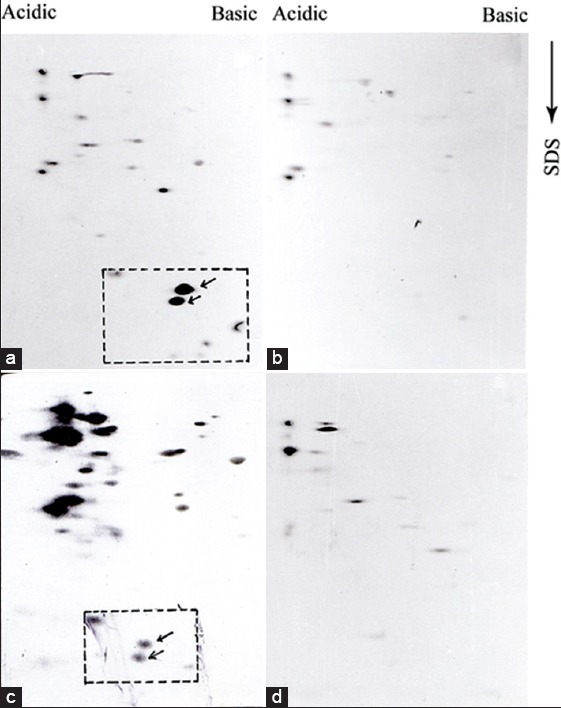
Figure 2.

A distinct difference is observed in immunoreactive protein pattern of pathogenic and nonpathogenic promastigote proteins: Protein extracts (~100 μg) of pathogenic (a and b) and nonpathogenic (c and d) promastigotes were analyzed by two-dimensional polyacrylamide gel electrophoresis and western blotted with virulent antisera (a and d) and avirulent sera (b and c) at 1:20 dilution. The low molecular weight proteins are enclosed with rectangle with arrows
