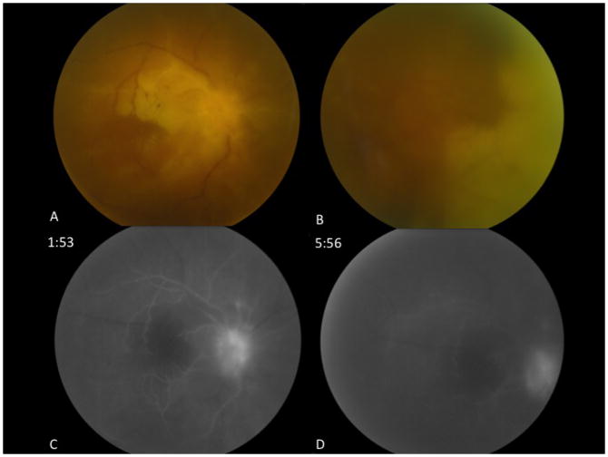
Figure 1.
Fundus photographs and fluorescein angiogram of the right eye. Fundus photograph of the posterior pole of the right eye shows a pale optic nerve, sclerotic retinal vessels and retinal whitening involving the macula (A). Confluent retinal whitening with obliteration of the vessels is observed nasally (B). Fluorescein angiogram of the right eye shows extremely delayed retinal arterial filling at 1:53 (C) with incomplete filling at 5:56 and late leakage of the optic disc (D).

