
FIGURE 3.
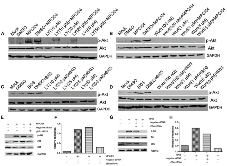
Role of PI3K in Akt phosphorylation. Serum-starved A549 cells were pre-incubated with LY294002 (LY; 10–50 μM), wortmannin (Wort; 100 nM–1 μM) or DMSO (0.4%, v/v) for 1 h and subsequently infected with MPC/04 (A,B) or B/03 (C,D) at a MOI of 5. A mock-infected lane was included in each panel as a negative control. (E,G) A549 cells were transfected with 15 pmol/well negative control (Negative siRNA) or p85α siRNA. 24 h after transfection, cells were infected with MPC/04 (E) or B/03 (G) at a MOI of 5. Cell lysates were collected at 30 min p.i. and separated by SDS-PAGE. In all assays, phosphorylated Akt (Ser473) was detected by Western blotting. Equal protein loading was verified using total Akt and GAPDH on the same membranes. The levels of p85α were determined by Western blot analysis using an anti-p85α antibody. (F,H) Quantification of relative p-Akt band intensities to Akt in (E) and (G). The results were confirmed in three independent experiments.