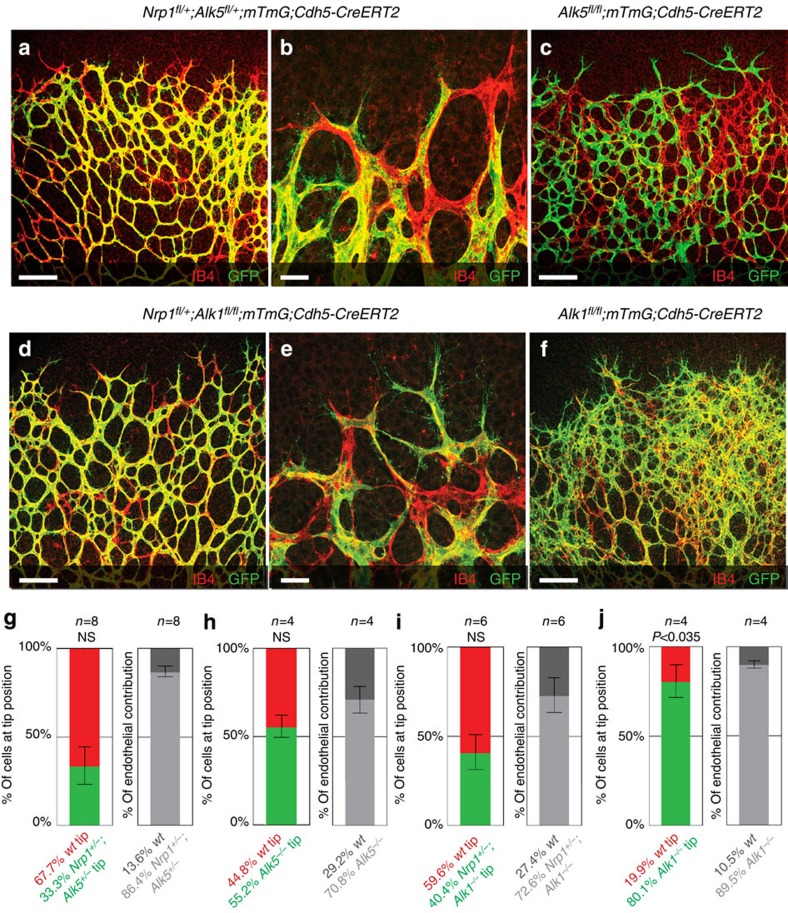
Figure 7. Inhibition of Alk5 and Alk1 rescues the Nrp1-deficient sprouting defect.
Retinas of Nrp1fl/+; Alk5fl/+; mTmG; Cdh5-CreERT2 mice (a,b), Alk5fl/fl; mTmG; Cdh5-CreERT2 mice (c), Nrp1fl/+; Alk1fl/fl; mTmG; Cdh5-CreERT2 mice (d,e) or Alk1fl/fl; mTmG; Cdh5-CreERT2 mice (f) injected with 30 μg tamoxifen at P1; retinas were assayed P5. Unrecombined wt cells are labelled with Isolectin-B4 only; recombined cells express GFP. Magnification of a is shown in b, magnification of d is shown in e; scale bar, 100 μm (a,c,d,f), 20 μm (b,e). Quantification of recombined Nrp1fl/+; Alk5fl/+ (g), Alk5fl/fl (h), Nrp1fl/+; Alk1fl/fl (i) or Alk1fl/fl (j) cells at the tip, normalized to overall contribution of cells to the endothelium. Statistical significance was determined by comparing the proportion of deficient (green) cells at the tip with the total proportion of deficient (green) cells; P=NS (0.0536) (g), P=NS (0.7576) (h), P=NS (0.275) (i), P<0.0358 (j). n=number of retinas analysed; n=8 (g), n=4 (h), n=6 (i) and n=4 (j); values represent mean±s.e.m. Statistical significance was assessed using a Student's unpaired t-test. NS, not significant.

