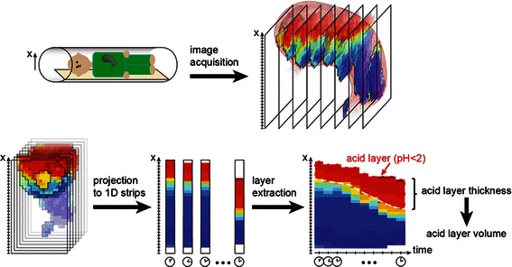
Fig. 3.

Schematic of post-processing steps for the quantification of acid layer thickness and volume from MR images. Gastric secretion accumulated predominantly on the meal surface and a secretion concentration gradient was observed from the surface of the meal into the test meal along the direction of gravity (i.e. the right side of the subject lying in the right lateral position) [10, 13]. The x-axis of the image data was set anti-parallel to the direction of gravity. Gastric content was sliced along the x-axis using a slice thickness of 1 mm and corresponding %secretion values were averaged along the other two Cartesian coordinates. This resulted in a 1D projection of mean %secretion values along the x-axis that was computed for each gastric secretion scan and stacked together over time to allow visualization of the formation of the gastric secretion layer (‘layer-graphs’). In the layer-graphs, the layer thickness was defined as the distance from the meal surface to the x coordinates having a threshold value of ≥70 % secretion. Layer volume (LV) was calculated by summing all pixels above this threshold
