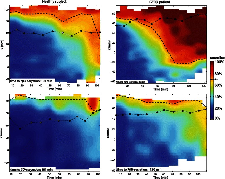
Fig. 7.

Layer graphs including EGJ position and time to Layer formation. Layer graphs of a healthy subject (left) and a GERD patient (right) under placebo (top) and PPI therapy (bottom). The vertical axis represents the MR image x-axis, which is aligned along the direction of gravity. The colours code the average %secretion. The threshold level of the layer (i.e. ≥70 % secretion) and the position of the EGJ are indicated as white dashed and black dotted line, respectively
