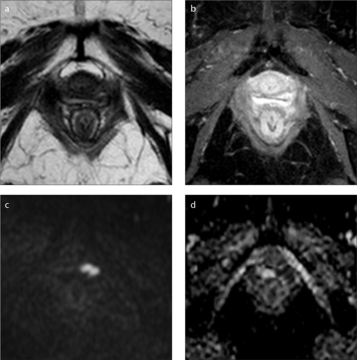
Figure 1.
a–d. MRI in the axial plane of the pelvis denoting vaginal recurrence of endometrioid adenocarcinoma of the cervix (initial stage IIB) treated with chemoradiotherapy. On T2-weighted (a) and dynamic contrast-enhanced (DCE) (b) MRI sequences, no obvious lesion was identified. On diffusion-weighted imaging (DWI) with b=1000 s/mm2 (c), the tumor is clearly defined with a hypersignal and low signal on the apparent diffusion coefficient (ADC) map (d).

