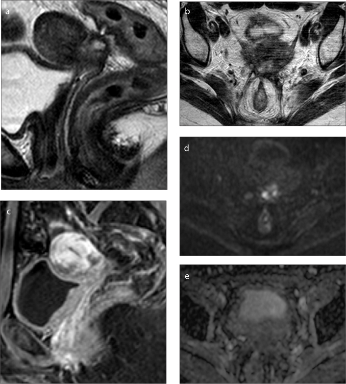
Figure 2.
a–e. Pelvic MRI of a 42-year-old patient who underwent chemoradiotherapy for cervical cancer that ended one year ago (initial stage IIB). On sagittal (a) and axial (b) T2-weighted images an irregular cervical nodular lesion is identified with necrotic center and ill-defined intermediate signal on the periphery. On sagittal DCE MRI (c) an early heterogeneous peripheral enhancement is seen. On axial DWI with b=1000 s/mm2 (d) the lesion shows diffuse hyperintense signal, mainly due to central necrosis and there is no peripheral low signal on the correspondent ADC map (e) denoting T2 shine-through effect, without evidence of restricted diffusion. Histopathology examination revealed only inflammatory changes.

