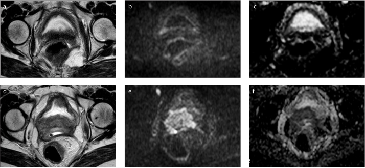
Figure 3.
a–f. Pelvic MRI of a 55-year-old patient who underwent chemoradiotherapy for cervical cancer (initial stage IIIB) with central recurrent disease after treatment. On axial T2-weighted image performed four years after treatment (a), no lesion was promptly identifiable, although DWI with b=1000 (b) showed a small focal area of hyperintensity on the right posterior bladder wall with a discrete hyposignal on the ADC map (c) that was not promptly considered relevant by the clinicians. After this examination, a targeted biopsy was performed confirming recurrence of cervical squamous cell carcinoma (this first evaluation was the one included in our study). The patient refused treatment for personal reasons and one year later, a hyperintense heterogeneous cervical mass with right posterior bladder invasion is readily visible on T2-weighted image (d) along with a high signal on DWI with b=1000 s/mm2 (e) and a low signal on the ADC map (f).

