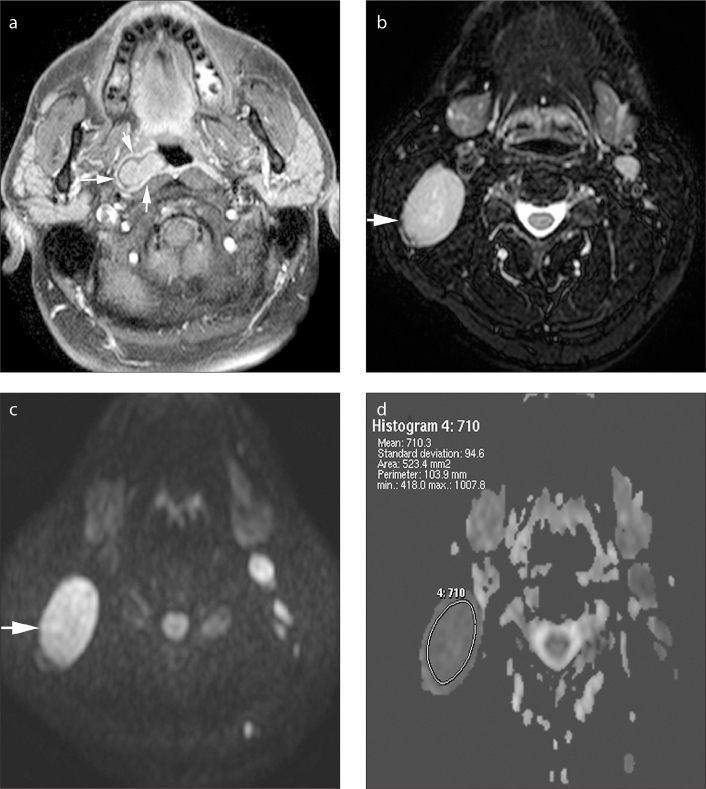
Figure 1.
a–d. Axial gadolinium-enhanced fat-suppressed T1-weighted image (a) in a 43-year-old male patient shows carcinoma of the nasopharynx on the right (arrows). Axial fat-suppressed T2-weighted image (b) demonstrates an enlarged, homogeneous metastatic lymph node at level 2, on the right (arrow). The lymph node is hyperintense on b=1000 s/mm2 DWI (c), and it appears as a hypointense lesion on the ADC map (d). The ADC value is 0.71×10⁻³ mm2/s.

