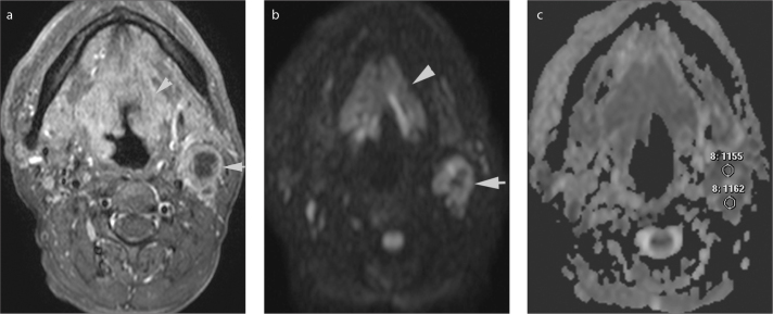
Figure 3.
a–c. Axial gadolinium-enhanced fat-suppressed T1-weighted image (a) in a 53-year-old male shows a squamous cell carcinoma of the supraglottic larynx extending cranially to the base of the tounge (arrowhead). There is a necrotic metastatic lypmh node at level 2, on the left (arrow). On b=1000 s/mm2 DWI (b) the lymph node was hyperintense with a central hypointense region that represents necrosis. On the ADC map (c) multiple small, uniform, round region of interests were placed on solid areas (two of them shown) and mean ADC was selected for statistical analyses.

