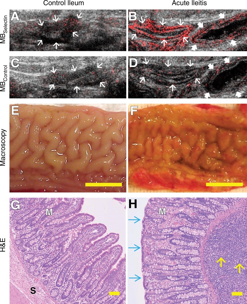
Figure 3:
A–D, Representative transverse US molecular images of control ileum and acutely inflamed terminal ileum imaged by using both MBSelectin and MBControl show background signal (arrows) only in, A, control ileum but increased signal in the bowel wall (thin arrows) in, B, severely inflamed bowel after injection of MBSelectin, while injection of MBControl resulted in background signal (thin and thick arrows) in both, C, control and, D, inflamed bowel. Note that an adjacent bowel segment with mild inflammation (thick arrows in B and D ) was also scanned and showed less imaging signal than did the severely inflamed segment. E, F, Macroscopic views of the lumen of the ileum show intact anatomy in, E, normal ileum and red and thickened mucosa in, F, acute ileitis. Scale bar = 1 cm. G, H, Photomicrographs of corresponding ileum tissue slices stained with hematoxylin-eosin (H&E) show normal mucosal (M) and submucosal (S) microanatomy in, G, control terminal ileum and severe inflammation in, H, acute ileitis with alteration of ileal microanatomy, including the presence of blunt and thicker villi (blue arrows) and expansion of the lamina propria and submucosa with a large number of neutrophils (yellow arrows). Scale bar = 100 µm.

