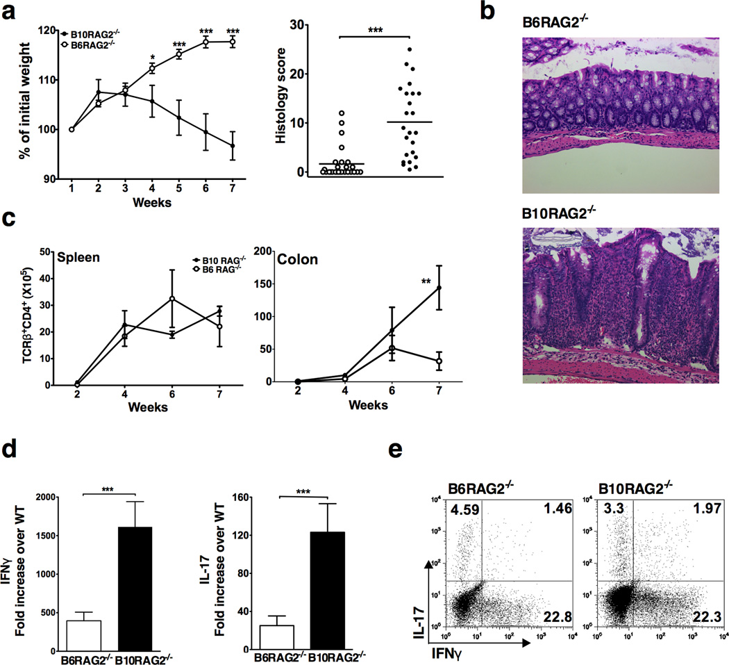
Figure 1.
B10-RAG2−/− recipients are more susceptible to T cell transfer colitis due to enhanced colonic accumulation of pathogenic T cells. (a) Weight change (Left panel) and colonic histology scores (right panel) at study end-point (7 weeks) following T cell adoptive transfer to B6-RAG2−/− and B10-RAG2−/− mice. Data shown are mean ± SD of 25 mice per group and representative of at least 3 independent experiments with 25–30 mice per group in each. (b) Representative colon histology in B6-RAG2−/− (score=2) and B10-RAG2−/− (score=25) mice at the study end-point (H&E-staining with original magnification 40X). (c) Total numbers of CD4+TCRβ+ cells isolated from the indicated organs at the indicated time points after T cell transfer. Data are mean ± SD of 5 mice per group per time point and representative of at least 3 independent experiments showing similar results with 20 mice per group in each experiment. (d) Real time RT-PCR analysis of IFNγ and IL-17 gene expression in the colonic tissue at the study end-point. Data are generated as mean fold increase ± SD, normalized to GAPDH (2ΔΔCt) of 38 and 33 animals per group in duplicate from 2 independent experiments. (e) Cytokine production by CD4+TCRβ+ lymphocytes isolated from the colonic lamina propria of B6 and B10-RAG2−/− mice at the study end-point and stimulated ex vivo for 6 hours in the presence of PMA/Ionomycin. Plots are representative of at least 3 independent experiments with at least 20 pooled colons per group in each experiment. Asterisks indicate statistically significant difference between the two strains (**P<0.01, ***P<0.001).

