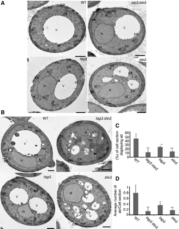
Lack of lipid droplets inhibits starvation-induced formation of autophagosomes
- A, B WT (SCY62), tagΔ (H1226), steΔ (H1112), and tagΔsteΔ (H1246) cells were grown to an exponential phase in rich medium and were then processed for electron microscopy (A) or starved in SD-N for 2 h, prior to electron microscopy analysis (B). Scale bars, 500 nm. CW, cell wall; ER, endoplasmic reticulum; M, mitochondrion; V, vacuole; SD-N, nitrogen starvation medium; WT, wild type. Arrows highlight homotypic membrane fusion of a vacuolar remnant. Arrowheads point to proliferating ER.
- C, D Average number of autophagic bodies per cell section (C) and percentages of cell sections displaying autophagic bodies in the vacuolar lumen (D) were determined by counting 100 randomly selected cell profiles. Error bars represent standard deviations from counting of the three grids. *P < 0.05, **P < 0.01 (Student’s t-test). WT, wild type.
