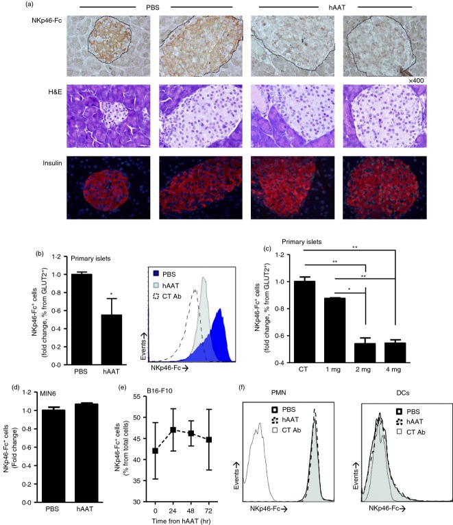
Figure 3.
Membrane-associated NKp46 ligand expression levels on pancreatic β-cells. DBA/2 mice were treated with human α1-antitrypsin (hAAT (2 mg per animal) or PBS for 3 days (n = 5 per group). (a) Representative histological images of immunohistochemical staining using soluble NKp46-Fc and horseradish peroxidase-conjugated anti-NKp46-Fc antibody (top), haematoxylin & eosin staining (middle) and anti-insulin immunofluorescence staining (bottom). Islet borders are demarcated by the dashed line. (b, c) NKp46-Fc+ cells derived from primary islets isolated from hAAT-treated (indicated doses) or PBS-treated DBA/2 mice (n = 5 per group). Fold change from PBS group; gated to GLUT2+ cells. Representative overlay is shown. (d) NKp46-Fc+ MIN-6 cells after hAAT treatment (0·5 mg/ml) for 72 hr. (e) NKp46-Fc+ B16-F10 cells after hAAT treatment (0·5 mg/ml) for 24, 48 and 72 hr. (f) Peripheral blood dendritic cells and polymorphonuclear cells were isolated from DBA/2 mice treated with 2 mg hAAT or PBS (n = 3 per group) and stained for NKp46-Fc. Data are representative of two to four experimental repeats. Mean ± SEM, *P < 0·05, **P < 0·01.

