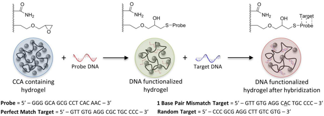
Figure 1.
Schematic of hydrogel functionalization with “probe” DNA and subsequent hybridization of “target” DNA strands. Color changes in the optically diffracting hydrogel are representative of those observed upon functionalization and hybridization due to changes in the lattice spacing of the encapsulated CCA. The sequences of the probe and target DNA strands that were used are shown below the schematic.

