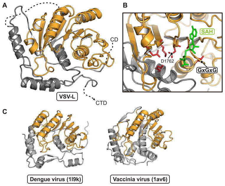
Figure 6. Methyl transferase domain.
(A) Structure of the methyl transferase: residues 1598–1892 in ribbon representation. The consensus fold of the S-adenosyl methionine-dependent methyl transferase subdomain is in orange. The N-terminal and C-terminal regions, in gray.
(B) Close-up of the active site. The SAM/SAH binding-site motif, GxGxG, is between β1 and αA. An SAH molecule (green) is derived from a superposition of its complex with dengue virus NS5 MT (PDB 1l9k). Residues that participate in the methyl transferase activity are in red.
(C) Comparison of VSV-L MT domain with other viral AdoMet-dependent methyl transferases. Structures of dengue virus NS5 MT (PDB 1l9k; residues 7–267) and vaccinia virus VP39 MT (PDB 1av6; residues 3–297) are shown in the same orientation and color scheme as in (A).
See also Figure S5.

