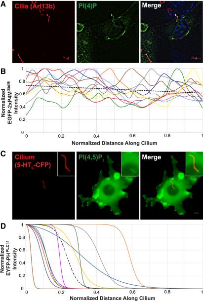
Fig. 1. PI(4)P and PI(4,5)P2 are present in distinct ciliary compartments.
(A) IMCD3 cells transfected with EGFP-2×P4MSidM, a PI(4)P sensor, were stained with antibodies against the ciliary protein Arl13b (red) and EGFP (green). Nuclei were marked by DAPI (blue). (B) Normalized EGFP-2×P4MSidM intensity for ten IMCD3 cilia was plotted against normalized distance along the cilium. The black dotted line is the linear regression of all cilia. (C) Cilia of live NIH-3T3 cells were visualized by 5HT6-CFP fluorescence (false colored red). PHPLCd1- EYFP, a PI(4,5)P2 sensor, accumulated in the proximal ciliary region (false colored green). Scale bar, 5μm. (D) Normalized EYFP-PHPLCd1 intensity for eleven NIH-3T3 cilia was plotted against normalized distance along the cilium and the data fitted to sigmoidal curves. The black dotted line is the average of all curves.

