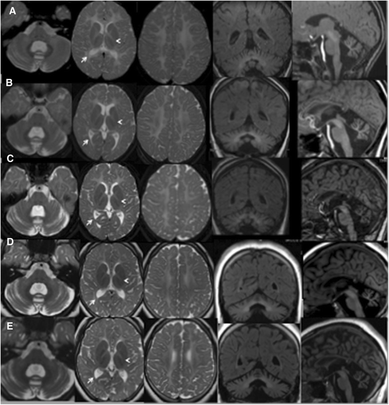
Fig. 1.

Hypomyelination with cerebellar atrophy: long term follow-up evaluation. a (7 years). b (10 year). c (13 years). d (15 years). e (19 years): Axial and coronal T2-weighted images show extensive cerebral white matter (WM) abnormalities with predominant involvement of the deep and subcortical WM; note the sparing of optic radiation (arrows), perirolandic WM and partial splenum corpus callosum. The head arrows (a-e), instead, indicate small hypointense dot in the posterior limb of the internal capsule. Mild abnormal hyperintensity involves the cerebellar WM. Sagittal T1-weighted images show a thin corpus callosum and shrunken cerebellar cortex with enlarged fissures. The pons is normal. At age of 13 years MRI (c) revealed a mildly increased cortical atrophy of cerebellar hemispheres that remained stable in the following MRI exams. No significative changes were observed during the yrs on WM abnormalies
