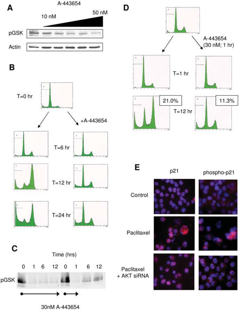
Figure 4.
A) Akt kinase assay following various doses of treatment with A-443654 in MIA-PaCa-2 cells demonstrates decreased phosphorylation of the target protein GSK, B) FACS analysis of MIA-PaCa-2 after the indicated times of treatment with paclitaxel (100 nM) in the absence or presence of the Akt inhibitor A-443654 (30 nM), C) Akt kinase assay following various times of treatment with A-443654 (continuous treatment on left; one hour treatment and media change on right) demonstrate mild recovery of Akt activity after 1 hr of Akt inhibition, D) FACS analysis of MIA-PaCa-2 cells in the absence (left) or presence (right) of the Akt inhibitor (30 nM) for 1 hour and then ongoing paclitaxel treatment (100 nM) with apoptotic fraction noted, E) Immunofluorescence for p21Cip/Waf1 and phospho-p21Cip/Waf1 in MIA-PaCa-2 cells following paclitaxel therapy in the absence (middle panel) or presence of Akt siRNa (bottom panel)

