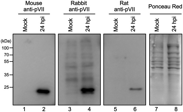
Fig 1. Western blotting using anti-protein VII antibodies.

U2OS cells were either mock-infected (lanes 1, 3, 5, and 7) or infected with Ad5 (lanes 2, 4, 6, and 8) and collected at 24 hpi. Cell lysates were prepared and subjected to 12% SDS-PAGE. Proteins were transferred to membranes and subjected to western blot analyses using mouse (lanes 1 and 2), rabbit (lanes 3 and 4), or rat anti-protein VII antibodies (lanes 5 and 6) or Ponceau Red staining as loading control (lanes 7 and 8).
