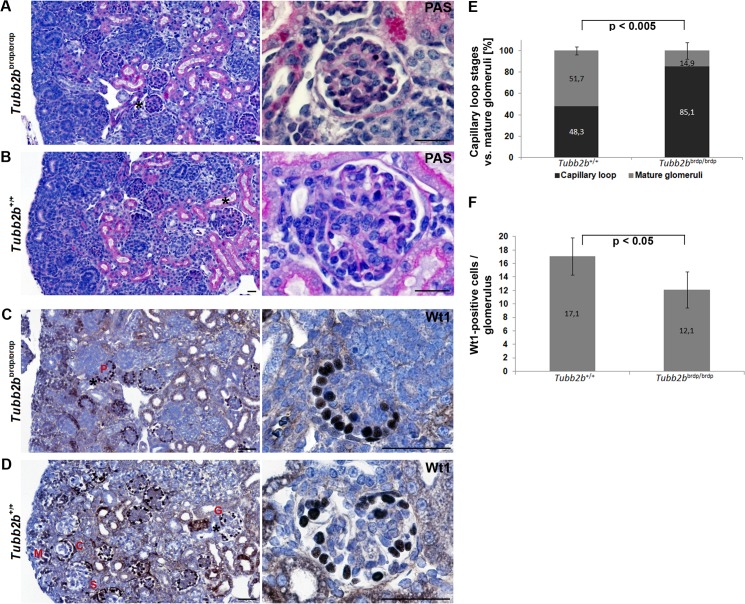
Fig 4. Periodic acid-Schiff and Wilm’s tumour 1 staining of wild type and Tubb2b brdp/brdp mouse kidneys (E18.5).
(A+B) Glomeruli were stained with periodic acid-Schiff to highlight basement membranes of glomerular capillary loops and tubular epithelium. (A) Tubb2b brdp/brdp mice often show a lack in glomerular tuft and capillary lumen development. (B) The capillary loops of the wild type glomeruli are well-defined and thin. Scale bars = 25 μm. (C+D) Wilm’s tumour 1 (Wt1) expression was detectable by immunohistochemistry (dark-brown nuclear staining results from 3,3'-diaminobenzidine). Nuclei were stained with hematoxylin (blue). (C) Tubb2b brdp/brdp kidneys show a specific Wt1 staining in podocytes (P) arranged in a string of pearls-like pattern at the periphery of the glomerulus, characteristic of an early developmental stage. (D) In the developing wild type kidney, Wt1 is expressed in mesenchymal cells that are starting the mesenchymal-to-epithelial transition (condensing metanephric mesenchyme (M)), in early epithelial structures (comma- (C) and S-shaped (S) bodies) and in fully differentiated epithelial cells (glomerular podocytes (G)). Black asterisks: Enlarged section areas on the right. Scale bars = 50 μm. (E) Percentage of capillary loop stages versus mature glomeruli in 3 mutant and 4 wild type animals. For each animal, 15 to 30 capillary loop stages / mature glomeruli were counted. Data are means ± SD. (F) Average number of Wt1-positive cells per glomerulus in 3 mutant and 4 wild type animals. 15 to 30 glomeruli / animal were counted. Data are means ± SD.

