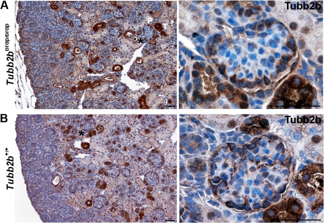
Fig 6. Tubb2b staining of wild type and Tubb2b brdp/brdp mouse kidneys (E18.5).
Immunohistochemical staining of mice kidneys (dark-brown Tubb2b staining results from 3,3'-diaminobenzidine. Nuclei were stained with hematoxylin (blue). (A) Tubb2b brdp/brdp kidneys show a specific cytoplasmic Tubb2b expression in tubuli (T) and in podocytes (P), confirming the developmental defects seen with the Wt1, Nphs2, Nphs1 and Synpo stainings. (B) Tubb2b expression in wild type kidneys is restricted to the mature podocytes. Interestingly in the developing wild type kidney Tubb2b is not expressed in the early developmental stages of maturing podocytes. Note, that in murine podocytes nuclear Tubb2b seems much less expressed compared to human kidneys. Black asterisks: Enlarged section areas on the right. Scale bars = 50 μm.

