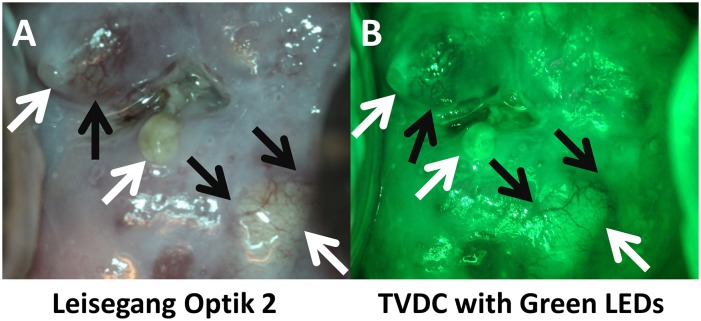
Fig 10. Comparison of green field images of a Nabothian cyst between the high-end digital colposcope and our POCkeT Colposcope.
The following image panel is of a biopsy confirmed normal cervix with Nabothian cyst (yellow and clear nodules) taken by the Leisegang Optik 2(AB) at 3.75X magnification and the 5.0MP POCkeT Colposcopes (CD) at 4X magnification. Black arrows indicate prominent vascular features and note the enhanced contrast with the “green field” illumination strategies by both systems. Both systems use White 5000K color temperature LEDs, while a green LED is use by the 5.0MP POCkeT Colposcope when compared to the short-pass Red filter used by the Leisegang Optik 2. Color correction was performed on the POCkeT Colposcope images using the color match function in Adobe Photoshop, GHI, JKL and original images in ABC,DEF. Here the illumination power threshold were set at 1% and 0.5% specular reflection level, respectively for white and green field illumination with the POCkeT Colposcope. We have now moved to a 0.5% and 0.05% specular reflection threshold for illumination power for all subsequent clinical cases.

