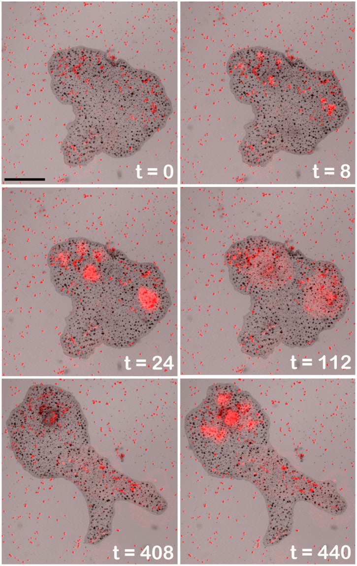
Fig 2. Trichoplax feeding on Rhodamonas salina microalgae.
Algae appear as tiny red specks on the substrate due to their content of fluorescent phycoerythrin. At 8 to 24 sec groups of algae under the paused Trichoplax precipitously release their contents apparent as contiguous domains of bright red phycoerythrin. The fluorescence has spread further and diminished at 112 sec. At 408 sec the animal has changed shape and a separate group of algae now covered by the paused Trichoplax release their contents at 440 sec. Merged transmitted light and fluorescence images (543 nm illumination) from a confocal microscope. Scale bar—200 μm.

