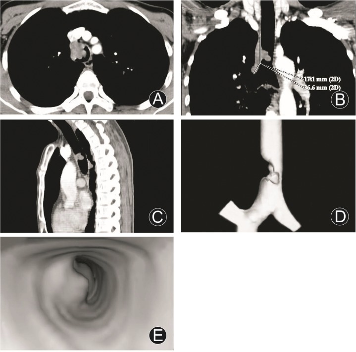
Fig 1. Squamous cell carcinoma of the lower trachea and involvement of right main bronchus.
MDCT axial image (A), MPR (coronal, B; sagittal, C), VR (D) and VB (E) images disclosed a wide based and irregular tumor, protruded into endolumen with severe eccentric lumenial stenosis, still homogeneous density and fairly obvious enhancement, extending to right main bronchus, and longitudinal involvement of 53.7 mm.

