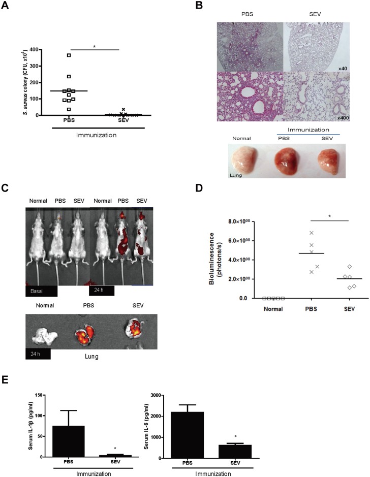
Fig 3. Efficacy of SEV vaccination on protection against pneumonia induced by sub-lethal dose of S. aureus.
For all figures, SEVs (5 μg) and sham (PBS) were injected intramuscularly to mice at weekly intervals for 3 weeks, and then sub-lethal dose (1 × 10⁸ CFU) of S. aureus was applied via the oropharyngeal route one week after the last immunization. Normal: PBS-immunized and PBS-challenged mice; PBS: PBS-immunized and S. aureus-challenged mice; SEV: SEV-immunized and S. aureus-challenged mice. (A) Colony forming unit (CFU) counts from lung of SEV- and sham (PBS)-immunized mice 24 h after the S. aureus challenge (n = 10, each group). (B) Histology (left panel) and gross image (right panel) of lung from SEV- and sham (PBS)-immunized mice after the sub-lethal dose of S. aureus challenge. (C) Distribution of S. aureus before and after SEV-immunization. Cy7-labeled S. aureus was applied via the oropharyngeal route to SEV- and sham (PBS)-immunized mice. Cy7 fluorescence of whole mouse (upper panel) or lung (lower panel) was acquired by IVIS spectrum 24 h after the S. aureus challenge. (D) Bioluminescence signal in the lung tissue after Cy7-labeled S. aureus administration. The amount of the bioluminescence signal (photons/s) in the lung tissue was measured by IVIS spectrum 24h after S. aureus challenge (n = 5, each group). (E) The levels of IL-β and IL-6 in serum of SEV- and sham (PBS)-immunized mice 24 h after the S. aureus challenge (n = 10, each group). * indicates p< 0.05 vs. PBS.

