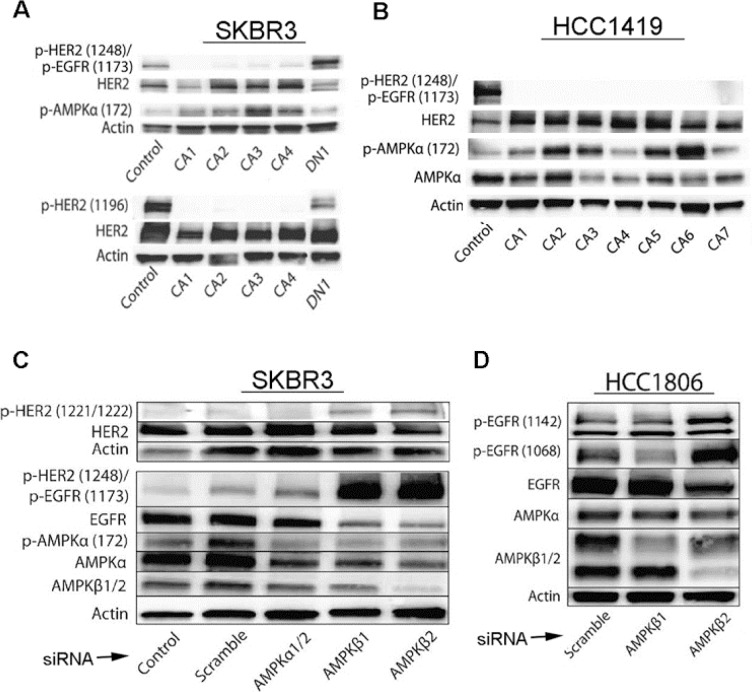
Figure 3. Effects of activated AMPK on phosphorylation of HER2 and EGFR is confirmed by genetic regulation of AMPK levels and activity.
A., B. SKBR3 and HCC1419 cells transfected with constitutively active (CA1 – CA4) AMPKα show decreased phosphorylation of HER2 (Y1248 and Y1196) and EGFR (Y1173), whereas cells transfected with dominant negative (DN) AMPKα show increased phosphorylation of HER2 (Y1248 and Y1196) and EGFR (Y1173). pAMPK was measured to monitor efficacy of CA and DN transfections. C. SKBR3 cells were transfected with AMPKα1/2, AMPKβ1, AMPKβ2, or scrambled control siRNA and three days after transfection, cell lysates were evaluated by immunoblot for HER2, pHER2(Y1248)/pEGFR(Y1173), EGFR, pAMPKα(172), AMPKα, AMPKβ1/2, and pHER2(Y1221-Y1222). Note increased levels of pHER2 (Y1248)/pEGFR (Y1173) and pHER2 (Y1221-Y1222) with AMPKβ1 or AMPKβ2 knockdown, while levels of AMPKα and AMPKβ are decreased by corresponding RNAi. D. HCC 1806 cells were transfected with AMPKβ1, AMPKβ2, or scrambled control siRNA, and three days after transfection, cell lysates were evaluated by immunoblot for p-EGFR (Y1142) and p-EGFR (Y1068). Note increased levels of EGFR protein phosphorylation at these sites with AMPK knockdown.

