
FIGURE 1. ABT-888 treatment and PARP-1 depletion sensitize MDA-MB-231 cells to doxo-induced apoptosis.
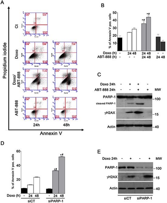
A. Apoptosis was analysed by FACS after treatment of MDA-MB-231 cells with 1 μM doxo and/or 0.5 μM ABT-888 for 24 and 48 h. Panels of a representative experiment are shown. B. Annexin V positive cells were counted in the right upper and lower squares. The diagram reports the percentage of Annexin V positive cells in untreated cells (black bar) and after treatment with 1 μM doxo (white bars), 1 μM doxo plus 0.5 μM ABT-888 (light gray bars) or ABT-888 alone (dark gray bars) at the indicated times in relation to total cells. Data represented are the mean+SEM of at least three independent experiments performed in duplicates. Comparisons were made with ANOVA/Turkey's test. *P < 0.05 compared to untreated cells; #P < 0.05 compared to cells treated with doxo at 24 h, 48 h respectively. C. Levels of cleaved PARP-1 (detected with mAb clone C2-10, Enzo Life Sciences) and γH2AX protein were measured by Western blot analyses in MDA-MB-231 cells treated for 24 h with 1 μM doxo and/or 0.5 μM ABT-888. D. Annexin V positive cells were counted in the right upper and lower squares. The diagram reports the percentage of Annexin V positive cells in siCT cells untreated (black bar) or treated with doxo (white bars) and in siPARP-1 cells untreated (black bar) or treated with doxo (light gray bars). Comparisons were made with ANOVA/Turkey's test. *P < 0.05 compared to untreated cell; #P < 0.05 compared to cells treated with doxo at 24 h, 48 h respectively. E. Levels of PARP-1 and γH2AX protein were measured by Western blot analyses in siCT MDA-MB-231 cells and in siPARP-1 MDA-MB-231cells treated for 24 h with 1 μM doxo.