
Figure 4. Fibulin-5 suppresses Wnt target gene expression by inhibiting ERK and GSK3β phosphorylation.
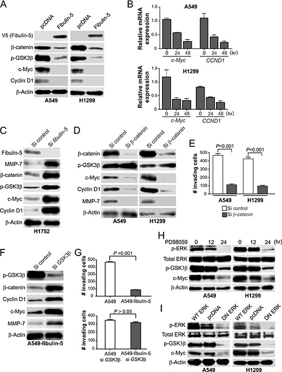
A. Western blot analysis of the indicated proteins in H1299 and A549 cells 24 hr after transfection with fibulin-5 or the control empty vector. B. Real-time RT-PCR analysis of c-Myc and CCND1 mRNA expression in H1299 and A549 cells at indicated time points after fibulin-5 transfection. Glyceraldehyde-3-phosphate dehydrogenase (GAPDH), a house-keeping gene, was used as an internal control. The results were normalized to the cells without fibulin-5 transfection (0 hr), which were defined as 1.0. C. Western blot analysis of the indicated proteins in H1752 cells at 48 hr after transfection with control or fibulin-5 siRNA. D. Western blot analysis of the indicated proteins in A549 and H1299 cells 36 hr after transfection with control or β-catenin siRNA. E. Matrigel invasion analysis of A549 and H1299 cells transfected with control or β-catenin siRNA as in (D). The results are the average of three independent experiments (P = 0.0001 for both si control and si β-catenin, Students' t test). F. Western blot analysis of the indicated proteins in stable fibulin-5-expressing A549 cells (A549-fibulin-5) 24 hr after transfection with control or GSK3β siRNA. G. Matrigel invasion analysis of the parental and stable fibulin-5-expressing A549 cells with or without GSK3β knockdown as in (F) (si control, P = 0.0001; si GSK3β, P = 0.1264, Student's t test). H. Western blot analysis of the indicated proteins in A549 cells and H1299 cells treated with 50 μM of the ERK inhibitor PD98059 for the indicated time. I. Western blot analysis of the indicated proteins in A549 cells and H1299 cells 24 hr after transfection with wild-type (WT) ERK, dominant negative (DN) ERK, or the control empty vector.