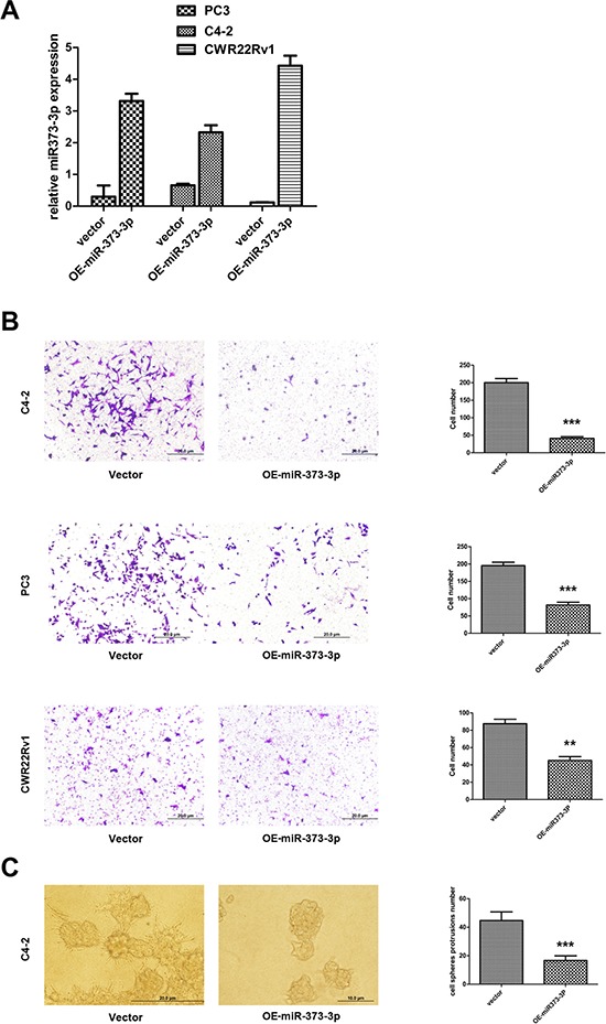
Figure 3. Effect of miR-373-3p on PCa cell invasion.

A–C. After stable lentivirus transfection, cells were seeded in a cell invasion chamber with the inner well coated with Matrigel and incubated for 24–36 h. A. Real-time PCR test expression (quantification) of miR-373-3p relative to U6 vector for stable overexpression (OE) of miR-373-3p in C4–2, PC3, and CWR22Rv1 cells. B. Invasion assay was performed in C4–2, PC3, CWR22Rv1 cells transfected with OE miR-373-3p or vector. Left: Representative microphotographs of invaded cells (magnification, 100 ×). Right: Quantitative analysis (cell numbers were counted in six randomly chosen microscopic fields per membrane). C. 3D spheroid invasion assay was performed in C4–2 cells transfected with OE-miR-373-3p or vector. Left: Representative microphotographs of cells (magnification, 200 ×). Right: Quantitative analysis (cell spheroid protrusions numbers were counted in six randomly chosen microscopic fields per membrane).*** P < 0.001, ** P < 0.01.
