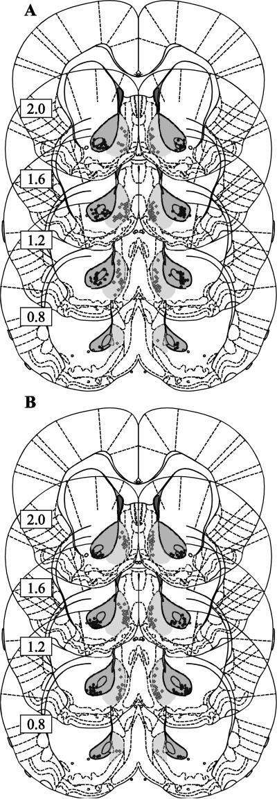
Figure 1. Histology.
The placement of the tip of the cannula is represented by a black circle for the core and a grey diamond for the shell for the animals in the first experiment (DAMGO, naltrexone, SCH23390 and raclopride experiments) in A and the second experiment (CTAP, naltrindole, nor-BNI) in B. Dark shading indicates the region that was acceptable for core cannula locations, and light shading indicates the same for the shell. Rats with one or both placements outside these regions were rejected from the analysis.

