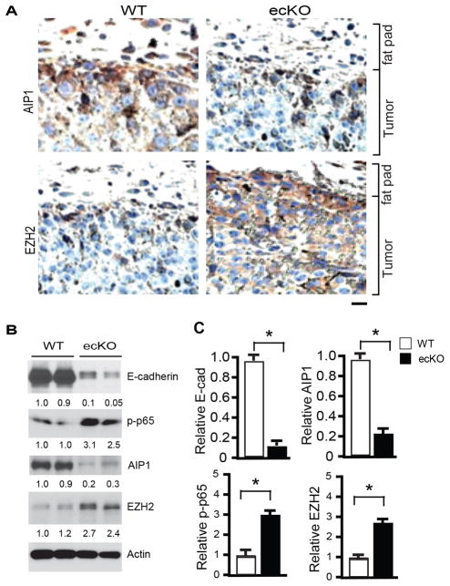
Figure 6. Tumor EMT is enhanced in AIP1-ecKO mice.
Mouse breast cancer cells (1×10⁶) were injected orthotopically into the fourth mammary gland of WT and AIP1-ecKO mice, and tumor along with surrounding mammary tissue were harvested at 2 weeks. A. AIP1 and EZH2 were detected by immunohistochemistry with respective antibodies as indicated. Tumor and surrounding fat fad are labeled. Scale bar: 50 μm. Representative images are shown from 5 sections per tumor and n=10 mice for each strain. B–C. Tumor EMT markers (E-cadherin, p-p65, AIP1 and EZH2) were determined by Immunoblotting with respective antibodies. Representative blots are shown in B from 2 of 10 mice for each strain with densitometry quantifications are shown below the blots. Quantifications are shown in C. n=10 mice per group. *, p<0.05.

