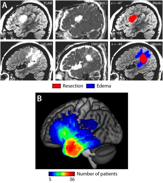
Figure 1.
Lesion masks and overlays. (A) Two example patients. Fluid Attenuated Inversion Recovery (FLAIR) and Diffusion Weighted Imaging (DWI) Apparent Diffusion Coefficient (ADC) images are shown. The first patient had no imaging abnormalities in addition to the resection; the second patient had edema adjacent to the resection. (B) Overlay of surgical sites in 110 patients.

