
Figure 1. SMB cells maintain key features of Shh-subtype MB.
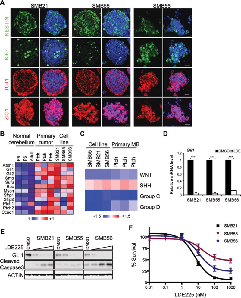
(A) Immunostaining of SMB cells with anti-Nestin, Ki67, Tuj1 Zic1. DAPI in blue. Scale bar: 20 μm.
(B) Microarray analysis reveals Shh-subtype signature in SMB lines and MB from Ptch+/− mice, compared to adult and P6 cerebellum.
(C) Expression of SMB cells, in vivo primary MB, P6 and adult cerebellum, analyzed using signature profiles of human MBs (WNT, SHH, Group C, Group D). Similarity to each subtype for each sample defined by “signature score” and normalized by normal mouse samples.
(D) Quantitative RT-PCR (qRT-PCR) analysis of Gli1 in SMB cells treated with DMSO or 1 μM LDE225 for 24 hrs. mean ± s.d., n = 3, *** p < 0.0001, Student’s t-test.
(E) Dose dependent inhibition of Shh signaling by LDE225 (0, 5, 50, 500 nM, 48 hrs). Shh signaling assessed by immunoblot for Gli1; Apoptosis assessed by cleaved Caspase3.
(F) Survival analysis of SMB cells treated with indicated LDE225 concentrations (72 hrs) (mean ± s.e.m., n = 6).