
Figure 7. RAS activation alters characteristics of Shh pathway-dependent tumors.
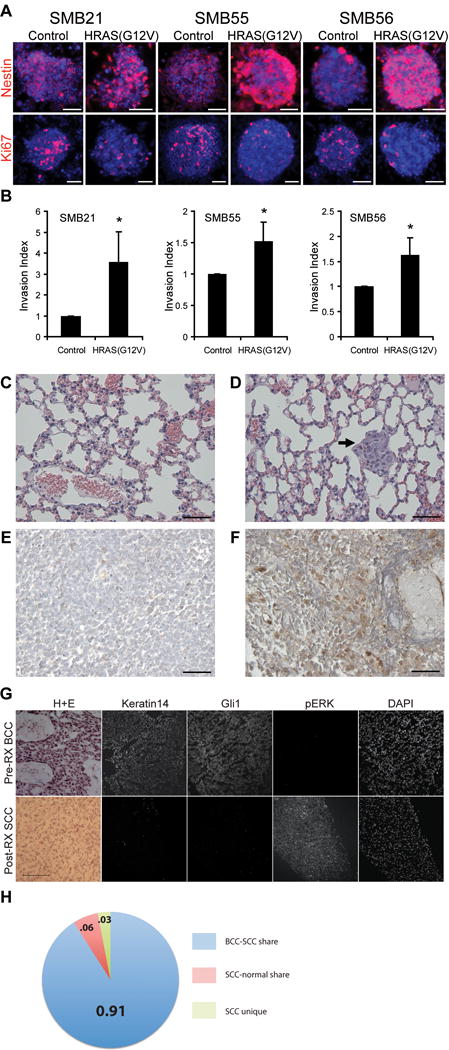
(A) SMB and SMB(HRAS) cells stained with anti- Nestin, Ki67; DAPI in blue. Scale bar: 50 μm.
(B) Cell invasion of SMB(HRAS) or SMB cells assessed using Matrigel-coated transwell apparatus. Invasion measured at 18 hrs, and normalized to SMB cells. (Mean ± s.d., n = 3, * p < 0.05, Student’s t-test)
(C–D) H&E-stained lung sections from animals engrafted with SMB (C) or SMB(HRAS) cells (D). Arrow: metastasis. Metastases detected in 2 of 9 HRAS mice; 0 out of 8 SMB parental mice. Scale bar: 50 μm.
(E–F) Phospho-ERK in primary and metastatic samples from human Shh-subtype MB. Phospho-ERK high in metastatic, frontal lobe tumor (F), but not in matched primary cerebellar tumor (E). Scale bar: 50 μm.
(G) Pre-treatment BCC with characteristic H+E, Keratin14, and Gli1 with an absence of phospho-ERK immunoreactivity. SCC tumor that developed at the same location after Vismodegib displayed a spindle-like morphology characteristic of SCC, and lacked keratin14 and Gli1 immunostaining, but was positive for phospho-ERK. Scale bar: 100 μm.
(H) Sequencing suggests shared lineage for post-treatment resistant SCC and pretreatment BCC. BCC and SCC share most genetic variations (n=1248, ratio=0.91); SCC-normal share (n=84, ratio=0.06); SCC unique (n=43, ratio=0.03).