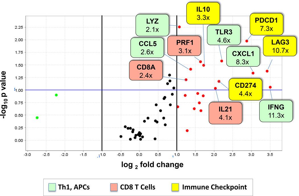
Figure 2. Functionally related genes are over-expressed in PD-L1+ melanomas.
RNA isolated from 6 PD-L1+ and 5 PD-L1(−) melanomas was assessed for expression of immune-related genes in a multiplex qRT-PCR assay. Results were normalized to PTPRC (CD45, a pan immune cell marker). Red and green dots represent genes over- and under-expressed by at least 2-fold, respectively, in PD-L1+ tumors. The horizontal blue line represents a p-value = 0.10. Numbers in colored boxes denote fold-change in gene expression. Over-expressed genes are associated with immunosuppressive molecules, activated CD8+ T cells, and antigen presenting cells. Additional information is provided in Supplementary Table S5. APC, antigen presenting cell; Th1, T helper 1.

