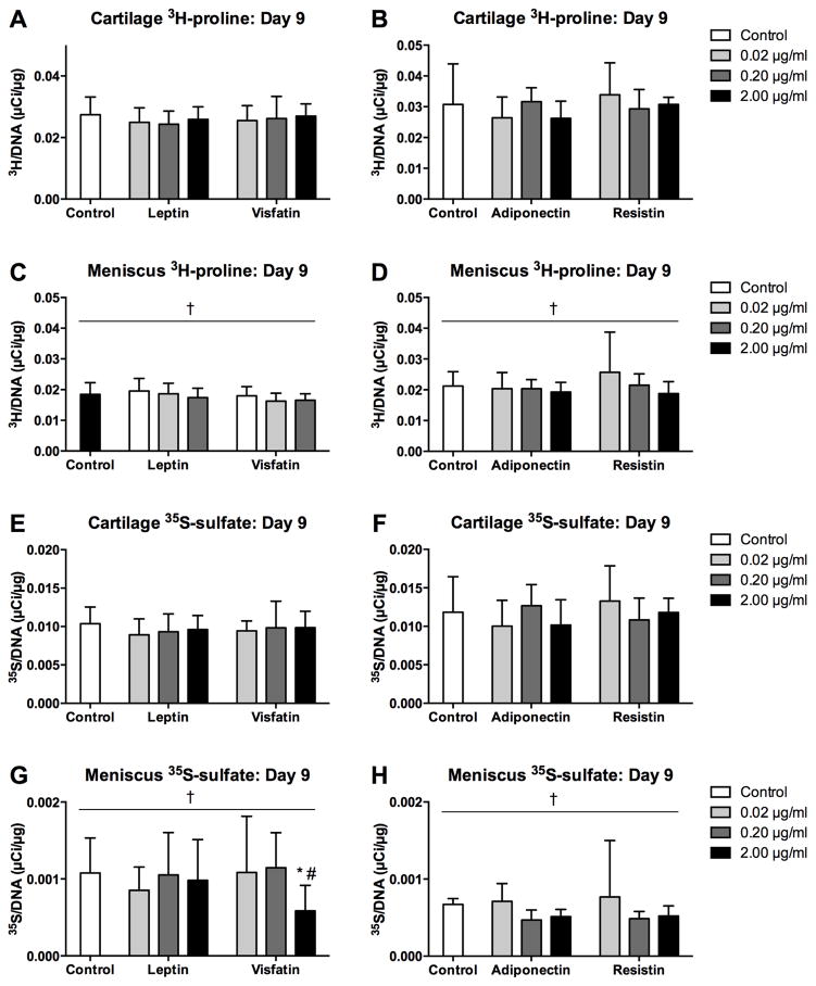
Figure 8. Effects of sustained adipokine exposure on incorporation rates.
Protein (3H-proline, A–D) and sGAG (35S-sulfate, E–H) incorporation rates of cartilage (A–B, E–F) and meniscus (C–D, G–H) explants over the final 22 hours of 9-day cultures with adipokines.. Note the difference in scaling between cartilage and meniscus. *p<0.05 relative to tissue-matched controls, #p<0.05 relative to 0.02 μg/ml treatment, †p<0.05 relative to cartilage tissue.

