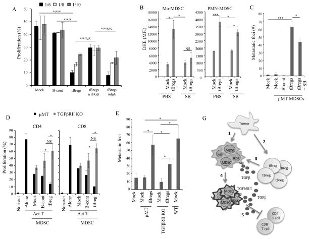
FIGURE 5. tBregs educate MDSC via TGFβ-TgfβR1/TgfβR2 axis.
4T1 cancer-bearing mouse μMT-MDSC (A–C) and TgfβR2 KO-MDSC (D,E) were stimulated in vitro with B-cont and tBregs with anti-TGFβ neutralizing antibody or mouse IgG (50 μg/ml) (A) in the presence or absence of SB431542 (20μM, SB, B and C). Then, MDSC were tested for T cell suppression as in Fig. 1G at indicated E:T ratio (A,D), or for expression of ROS (B), or induce metastases upon transfer into μMT mice (C,E). Prior to the transfer or use in suppression assays, MDSC were depleted of B cells. Y-axis shows DHE staining (MFI) in Mo-MDSC and PMN-MDSC (B), % T cell proliferation (gated in CD4+ and CD8+ T cells, A,D), and # of metastatic foci in the lungs of μMT mice (C,E) ± SEM in four-five mice per group experiments reproduced three times. (G) Summary: Tumor initiates an expansion of MDSC (1) and conversion of tBregs from naïve B cells (2). Cancer-activated B cells/tBregs use TGFβ (3) to induce TGFβ receptors on MDSC (4) and to up regulate ROS and NO production in MDSC. As a result, MDSC become fully suppressive for T cells and thereby pro-metastatic (5). *P<0.05, **P<0.01, ***P<0.001.

