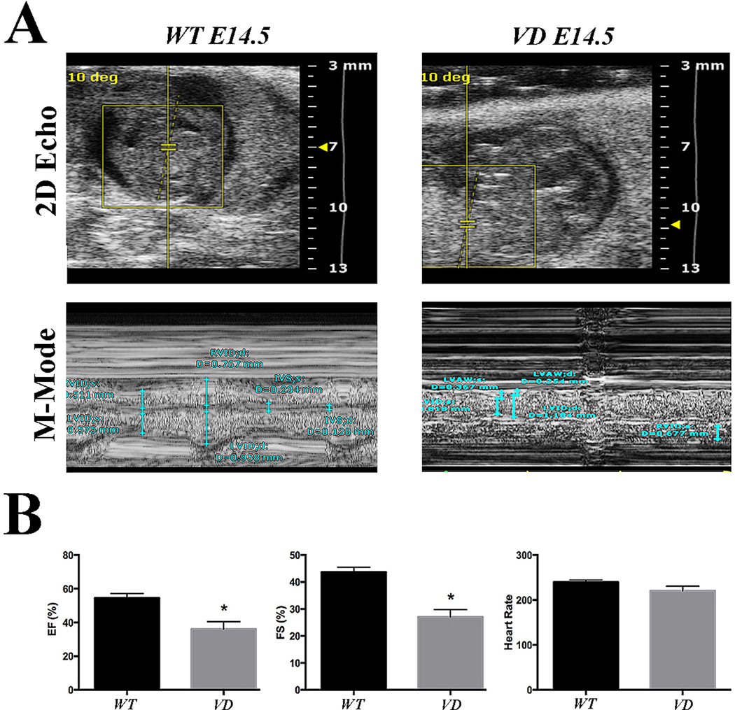
Figure 3. VD embryos reveal reduced cardiac function.
(A) Representative 2D images and M-mode analysis of E14.5 embryos. VD hearts show reduced systolic function (n=4). (B) Quantification of the ejection fraction (EF), fractional shortening (FS) and heart rate. The EF and FS are significantly reduced in the VD embryonic hearts (n=4). *p < 0.05

