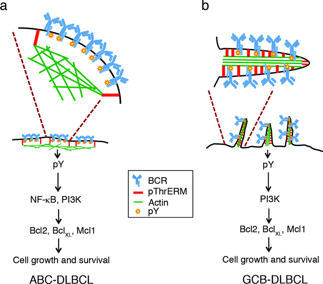
Figure 8. Model for regulation of BCR signaling by ERM proteins in DLBCL.
(A) In ABC-DLBCL with activating CD79 mutations, the pre-clustered BCRs are trapped within compartments supported by phosphorylated ERM proteins through linkage of plasma membrane and actin filaments. These BCR clusters continuously transduce NF-κB and PI3K pathway activation signals to promote cell growth. (B) In GCB-DLBCL, the BCRs are present in surface microvilli, whose backbone consists of phosphorylated ERM proteins and actin filaments. The BCRs transmit tonic signals through the PI3K pathway in this location. ERM inhibition disrupts both types of organization by interfering with plasma membrane-actin cytoskeletal linkage.

