
Figure 2.
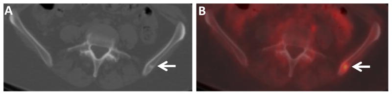
FDG-avid small sclerotic osseous metastasis in an IDC patient. Axial CT (A) and fused FDG PET/CT (B) images through the pelvis of a 56 year old woman with IDC demonstrate a small FDG-avid sclerotic osseous lesion in the left ilium (arrows). Similar small FDG-avid lesions were seen in the left scapula and left ischium (not shown). Osseous metastasis was confirmed by biopsy.