
Figure 3.
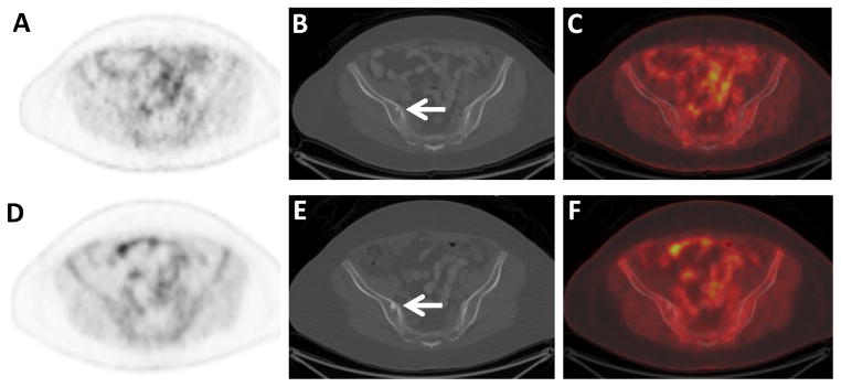
Non-FDG-avid sclerotic osseous metastases in an ILC patient. Axial FDG PET (A), CT (B), and fused FDG PET/CT (C) images through the pelvis of a 60 year old woman with ILC demonstrate a small non-FDG-avid sclerotic osseous lesion on CT (arrow). Additional small non-FDG-avid sclerotic osseous lesions were seen in the spine (not shown). Axial FDG PET (D), CT (E), and fused FDG PET/CT (F) images following adjuvant systemic therapy demonstrate increasing size of the osseous lesions, considered to be either flare response from treatment of osseous metastases or increasing metastases (arrow). Other sclerotic lesions had also increased in size (not shown).