Fig. 1.
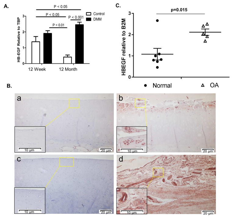
HB-EGF is increased in a mouse model of OA and in human OA cartilage. (A) OA was surgically-induced by destabilization of the medial meniscus (DMM) in 12 week-old (n=9 sham controls and n=9 DMM) and 12 month-old mice (n=9 sham controls and n=9 DMM). RNA was isolated from the knee joint tissue at 8 weeks after surgery. Samples pooled from 3 animals per group were used for real-time PCR normalized to TBP as a housekeeping gene. Results are the mean±sem of three independent RNA pools per condition. (B) Immunohistochemical staining for HB-EGF on human cartilage sections from a young normal donor (a, 45 year-old), OA patient (b, 90 year-old), old normal donor (c, 70 year-old) and young normal donor meniscus (d, 48 year-old). Colorimetric detection was with NovaRed substrate. Results are representative of staining from four different donors for each group (young normal, old normal, and OA). (C) Real-time PCR analysis for HB-EGF expression using RNA isolated directly from normal (n=7, avg age 53.1 years) and OA (n=5, avg. age 57.4 years) cartilage samples normalized to B2M expression as a control.

