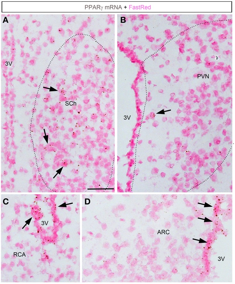
Figure 4.
Chromogenic detection of PPARγ in the mouse hypothalamus. (A) PPARγ hybridization signals (brown) in the suprachiasmatic nucleus. Moderate expression was seen in scattered cells. (B) PPARγ hybridization signals in the paraventricular hypothalamus were very limited. (C) PPARγ hybridization signals in the retrochiasmatic area are observed in the mediobasal portion of the 3rd ventricle, but virtually absent from adjacent neurons. (D) The mediobasal portion of the 3rd ventricle contained positive ependymal cells. Neurons in adjacent ARC showed very little expression. Minor adjustments in contrast, brightness and sharpness were made uniformly. Tissue was counterstained with Fast-Red (pink). Abbreviations: 3V, third ventricle; ARC, arcuate nucleus of the hypothalamus; PVN, paraventricular nucleus of the hypothalamus; RCA, retrochiasmatic area. Scale bar is 40 μm. Arrows indicate representative positive cells.

