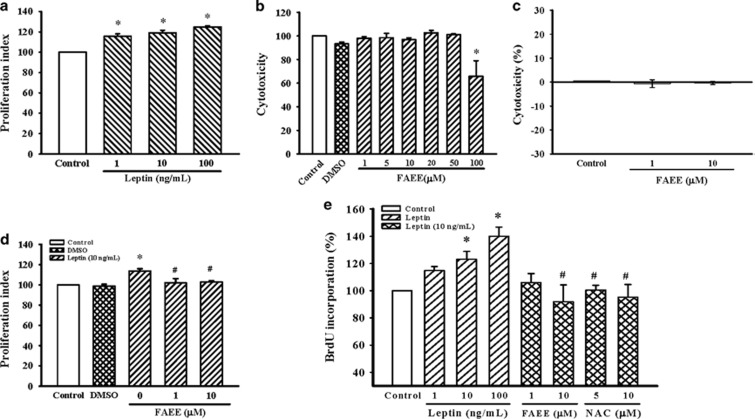
Figure 1.
Effect of FAEE on leptin-induced proliferation of A10 cells. (a, d) Growth-arrested cells were stimulated with leptin (1–100 ng ml−1) for 72 h in the presence or absence of FAEE (1 and 10 μM). Cell proliferation was assayed by the CellTiter 96A Queous One Solution kit. Relative proliferation activities were expression using untreated control cells as a standard. Data represent as mean±s.e. of six independent observation with different cell passages and on different days. *P<0.05 vs control; #P<0.05 vs leptin alone. (b, c) Cells were incubated with FAEE at increasing concentrations (1–100 μM) for 24 h; the toxic effects of FAEE were measure by the CellTiter 96A Queous One Solution kit and LDH cytotoxicity assay kit, respectively. Data represent as mean±s.e. of six independent observation with different cell passages and on different days. *P<0.05 vs Control. (e) DNA synthesis was measured by BrdU incorporation assay. Growth-arrested cells were stimulated with leptin (1–100 ng ml−1) for 72 h in the presence or absence of FAEE (1 and 10 μM) or NAC (5 and 10 μM). Data represent as mean±s.e. of six independent observation with different cell passages and on different days. *P<0.05 vs control; #P<0.05 vs leptin alone.

