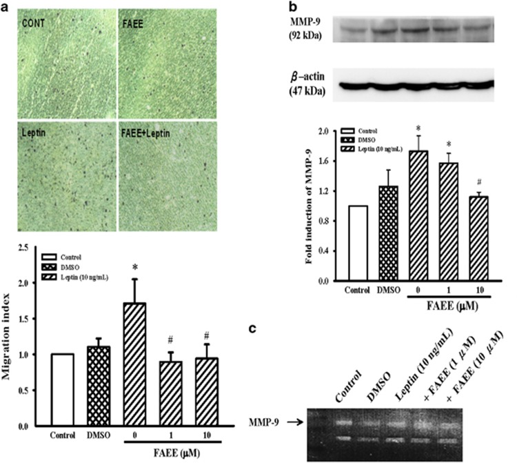
Figure 6.
Effects of FAEE on leptin-induced VSMC migration as well as MMP-9 expression in A10 cells. (a) VSMCs migration was examined using Transwell Permeable Support Culture Plate System. After incubation at 37 °C for 48 h, migrated cells were counted at × 200 magnification in five randomly chosen microscope fields per filter. Lower panel indicated the fold value of cell migration. Data represent as mean±s.e. of four independent observation with different cell passages and on different days. (b) Cells were stimulated with leptin (10 ng ml−1) for 3 h in the presence or absence of FAEE (1 and 10 μM). The cells were lysed and MMP-9 protein was analyzed by western blotting. The β-actin was used for normalization. After densitometric quantification, data represent as mean±s.e. of five independent observation with different cell passages and on different days. *P<0.05 vs control; #P<0.05 vs leptin alone. (c) Gelatin zymography analysis was performed with conditioned media collected from A10 cells cultured in the presence or absence of FAEE and leptin (10 ng ml−1).

