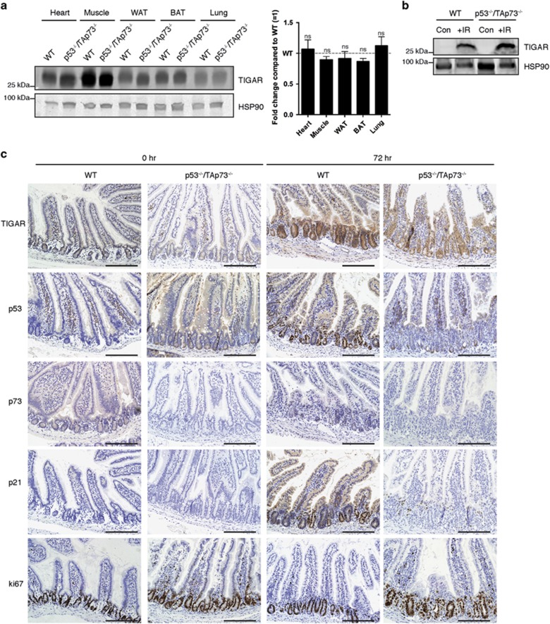
Figure 6.
TIGAR expression in p53- and TAp73-null animals. (a) Left: Western blot analysis of TIGAR protein expression in organs of wild-type (WT) and p53−/−TAp73−/− mice. Right: Graph represents quantification of western blots with fold change compared with WT. (b) Western blot analysis of TIGAR protein expression in small intestine tissue of WT and p53−/−TAp73−/− mice 72 h after 10 Gy IR. Tissues were harvested from one experiment. (c) Immunohistochemistry on small intestines from WT and p53−/−TAp73−/− animals 72 h after 10 Gy IR. Scale bar, 20 μm. Values represent mean±S.E.M. of two independent experiments unless otherwise indicated. NS, not significant; WAT, white adipose tissue; BAT, brown adipose tissue

