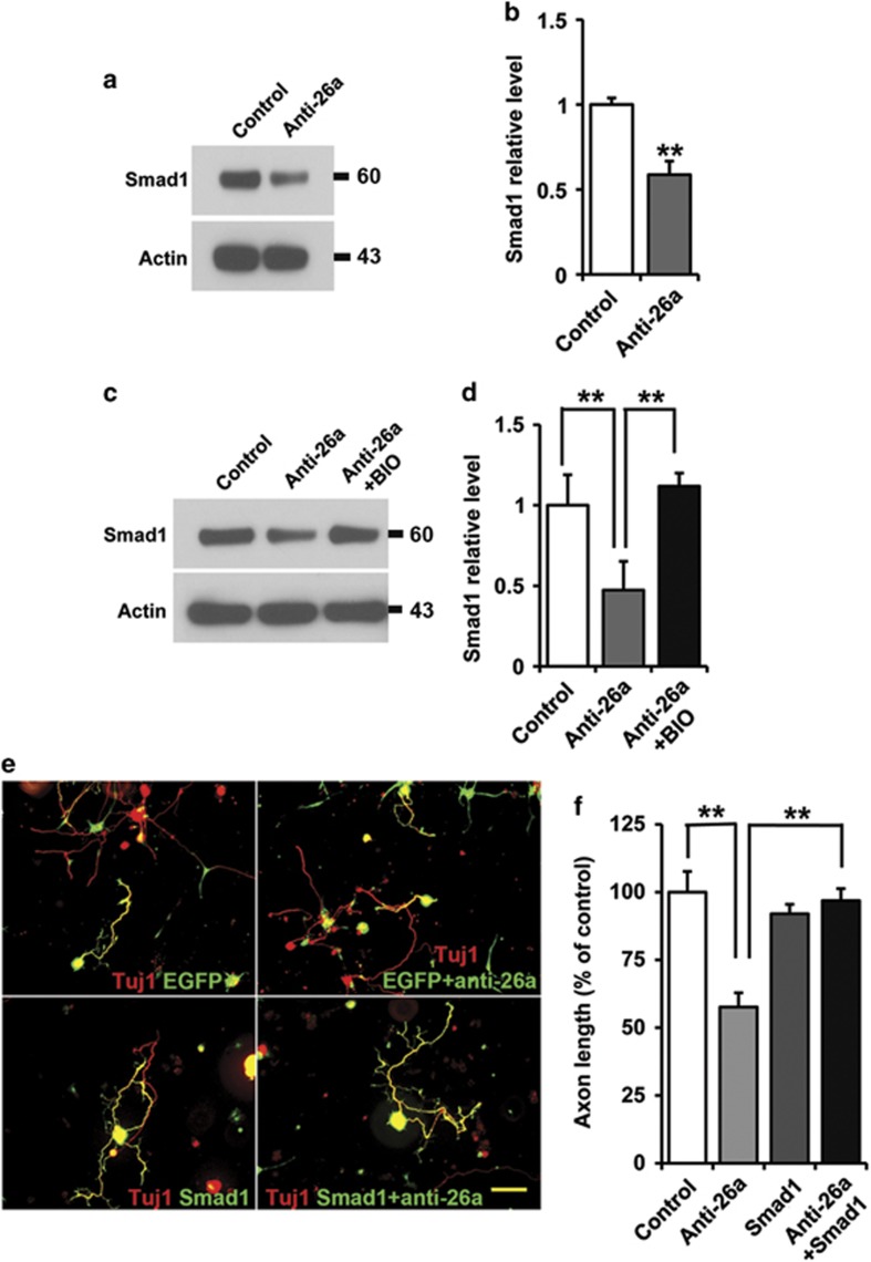
Figure 4.
MiR-26a-GSK3β regulate axon regeneration by controlling Smad1 expression. (a) Representative western blot images of Smad1 in cultured adult mouse sensory neurons 3 days after inhibition of miR-26a. (b) Quantification of Smad1 level (normalized to actin, n=3). (c) Representative western blot images of Smad1 in cultured adult mouse sensory neurons 3 days after inhibition of miR-26a with or without the treatment the GSK3 inhibitor BIO. (d) Quantification of Smad1 level (normalized to actin, n=3). (e) Representative images of cultured adult mouse sensory neurons expressing EGFP, EGFP+miR-26a inhibitor (anti-26a), Smad1 and Smad1+anti-26a. Scale bar=100 μm. (f) Quantification of the average length of the longest axons (normalized to the average length of the control axons, n=3). Bar graphs are shown as means±S.E.M. **P<0.005

