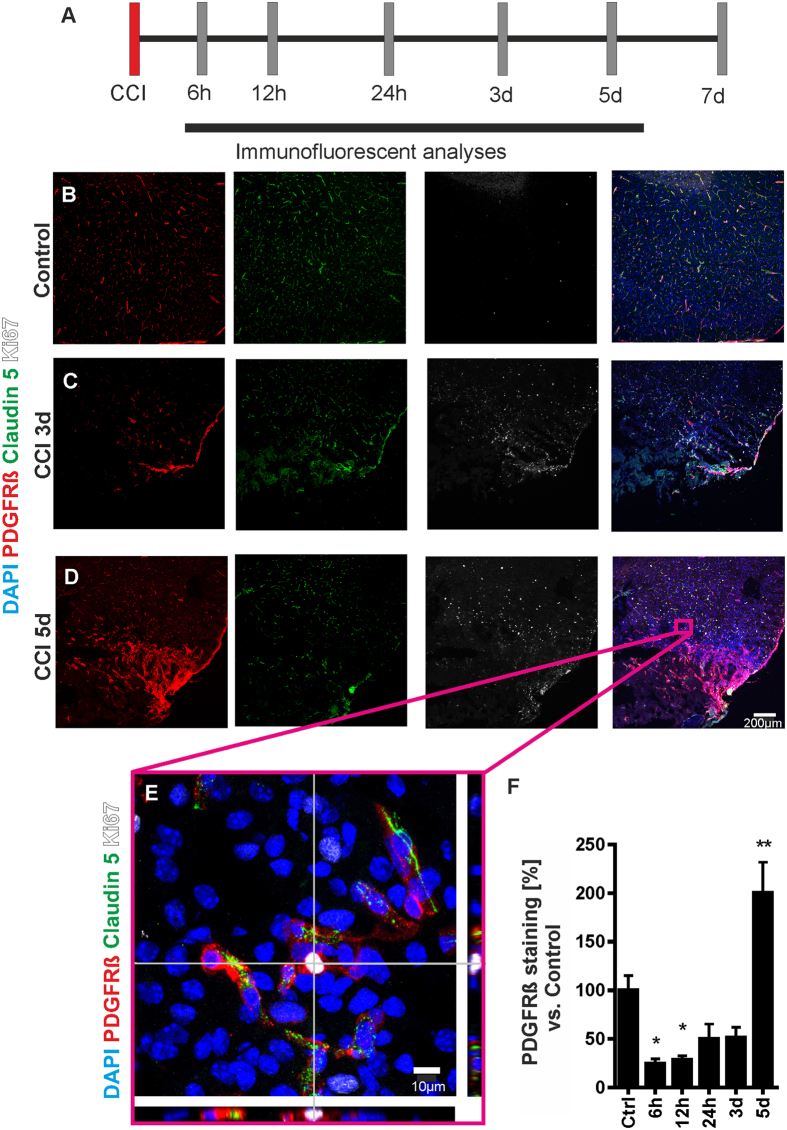
Figure 1. CCI results in pericontusional pericyte proliferation.
Tissue was collected for immunohistochemical and mRNA analyses at varying time points after induction of CCI (A). Under physiological conditions pericytes were located in a typical perivascular manner within the cerebral cortex (Claudin 5: green; PDGFRß: red). Only few cells were positive for Ki67, a marker for cell proliferation (B). Three days after CCI PDGFRß positive cells at the CCI border were associated with a strong increase of Ki67 immunoreactivity (C). Five days after CCI a prominent increase of PDGFRß expressing cells was detected (D). Panel E shows an orthogonal section view of a pericontusional pericyte positive for Ki67 that is not in contact with a microvessel. Quantification of PDGFRß staining demonstrates an initial loss of PDGFRß that is followed by a significant increase of PDGFRß 5 days after CCI (F). N = 5–6 animals per group; *P < 0.05; **P < 0.01.

